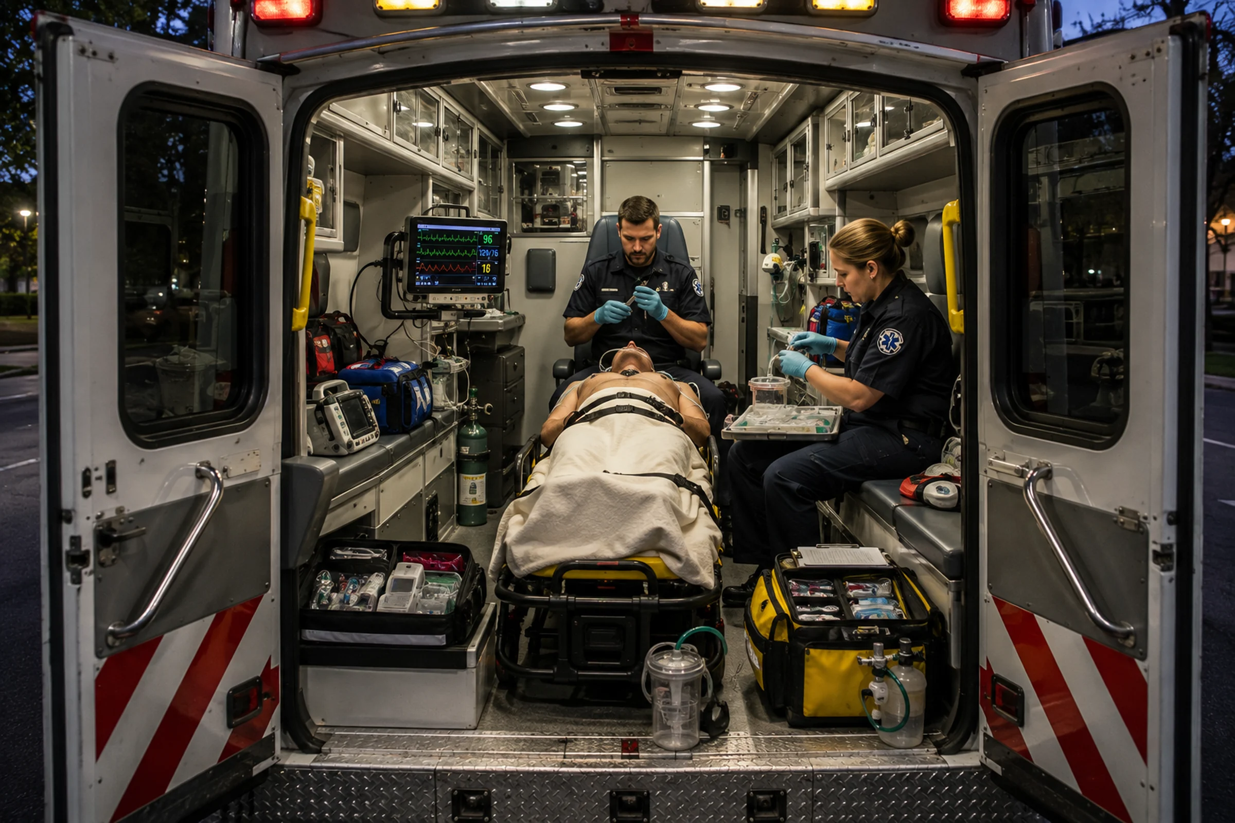
💡 Clinical note: This article is for EMS education and does not replace agency protocols, medical direction, or patient-specific clinical judgment.
The endotracheal tube passes through the cords.
The cuff inflates.
Waveform capnography appears.
The tube is secured.
Successful intubation.
Or was it?
For decades, EMS airway performance has often centered on one easily measured outcome:
Did the tube go in?
Modern airway management demands a more important question:
What happened to the patient while we were putting it in?
A successful endotracheal tube does not erase severe hypoxemia, profound hypotension, bradycardia or peri-intubation cardiac arrest.
And new national EMS research reinforces why airway management should be viewed as a physiologic resuscitation, not simply a procedural skill.
Looking Beyond Tube Placement
Researchers examining prehospital neuromuscular-blockade-assisted airway management used data from the 2023 National Emergency Medical Services Information System.
The analysis included adult 911 encounters involving neuromuscular-blockade-assisted airway management and excluded patients already in cardiac arrest.
The investigators were interested in physiologic adverse events surrounding airway management, including:
- hypoxemia,
- hypotension,
- bradycardia,
- and cardiac arrest.
That approach matters because airway quality cannot be adequately described by intubation success alone.
The patient can have a perfectly positioned endotracheal tube and still suffer major physiologic deterioration during the procedure.
Intubation Changes Physiology
Endotracheal intubation is not physiologically neutral.
Critically ill patients may already be maintaining life through compensatory mechanisms that are close to failure.
A spontaneously breathing patient generates negative intrathoracic pressure.
Sedation, neuromuscular blockade and positive-pressure ventilation change that relationship.
Positive pressure can reduce venous return.
Induction medications can alter vascular tone and cardiovascular performance.
Apnea eliminates the patient’s spontaneous ventilation.
A patient with limited oxygen reserve may desaturate rapidly.
A patient with shock may lose the compensatory mechanisms maintaining an already marginal blood pressure.
A severely acidotic patient who has been maintaining a high minute ventilation can deteriorate if post-intubation ventilation does not meet the physiologic demand that existed immediately before the procedure.
The airway may improve while the patient gets worse.
The Difficult Airway Is Not Always Anatomically Difficult
EMS airway training appropriately spends substantial time on anatomy.
Mouth opening.
Mallampati assessment.
Neck mobility.
Facial trauma.
Obesity.
Airway contamination.
Anatomically difficult airways deserve preparation.
But another category is equally important:
the physiologically difficult airway.
This is the patient whose anatomy may permit an easy first-pass tube but whose physiology gives the team very little margin for error.
Examples may include patients with:
- severe hypoxemia,
- shock,
- major hemorrhage,
- right-ventricular failure,
- severe metabolic acidosis,
- or profound respiratory failure.
The challenge is not necessarily seeing the cords.
The challenge is getting through the procedure without causing cardiovascular or respiratory collapse.
Preoxygenation Is Part of the Procedure
Preoxygenation should not be viewed as waiting time before intubation.
It is an intervention designed to increase the patient’s oxygen reserve before apnea.
The method needs to match the patient.
A cooperative patient with relatively preserved physiology may require a very different strategy from someone with severe shunt physiology, pulmonary edema or significant hypoxemic respiratory failure.
Teams should think beyond simply placing a nonrebreather on the patient and calling the airway prepared.
- Is oxygen saturation improving?
- Is the mask sealing?
- Would positive pressure improve preoxygenation?
- Does the patient tolerate positioning?
- Can apneic oxygenation be used?
- Is suction ready if contamination occurs?
The answer is not a single device.
The answer is an oxygenation strategy.
Resuscitate Before You Intubate
One of the most important concepts in modern emergency airway management is resuscitation before intubation when time and circumstances permit.
That does not mean delaying a necessary airway indefinitely.
It means recognizing modifiable risks before medications remove the patient’s compensatory physiology.
Consider the patient with profound hypotension.
If the patient is barely maintaining perfusion before induction, sedation and positive-pressure ventilation may worsen that instability.
Teams should identify and address reversible contributors when possible.
That may include hemorrhage control, appropriate fluid or blood-product resuscitation, vasoactive support when indicated, oxygenation and positioning.
The airway plan should be integrated into the resuscitation plan.
Not performed separately from it.
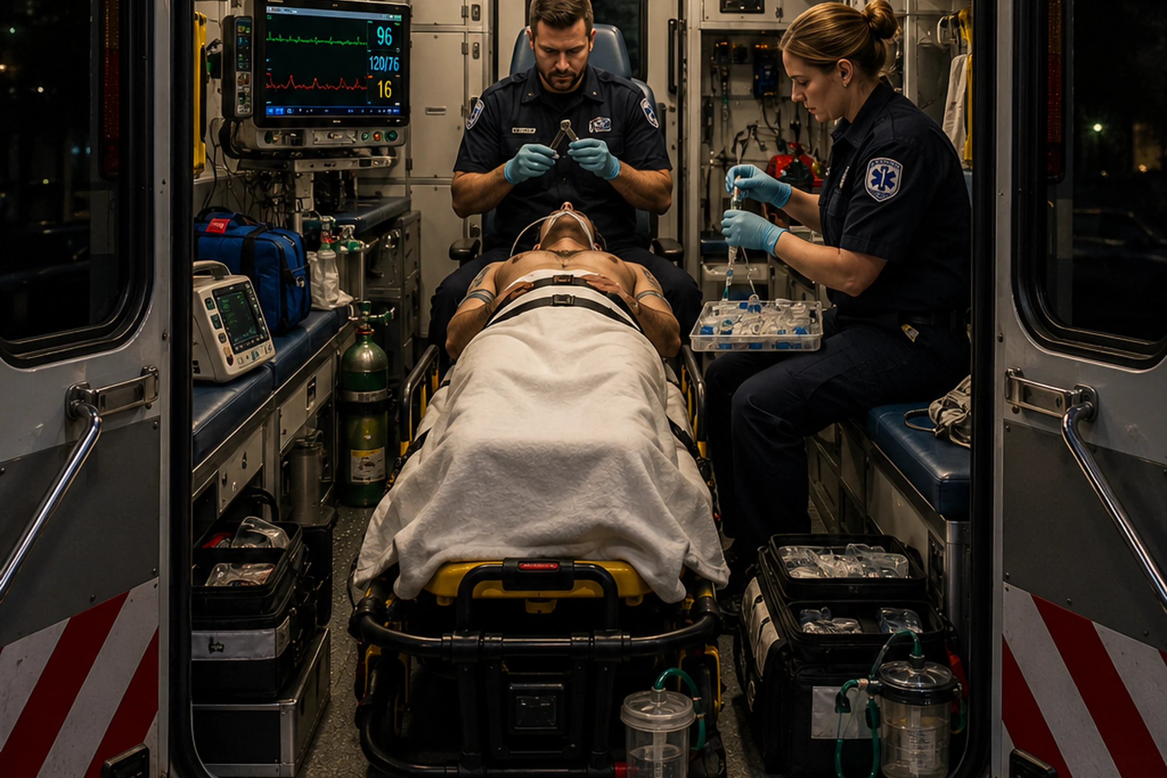
The Airway Checklist Should Include Physiology
Airway checklists often include equipment:
- laryngoscope,
- tube,
- stylet or bougie,
- suction,
- backup airway,
- capnography,
- medications.
Those are essential.
But the checklist should also force the team to evaluate the patient.
- What is the oxygen saturation?
- What is the blood pressure?
- Is the patient in shock?
- What is the anticipated post-intubation ventilation strategy?
- What is the failed-airway plan?
- Who is watching physiology during the attempt?
A good checklist should make it difficult for the team to become so focused on equipment that they forget the patient attached to it.
First-Pass Success Matters, but It Isn’t Everything
First-pass success remains an important airway metric.
Multiple attempts can increase the risk of hypoxemia, airway trauma, aspiration and physiologic deterioration.
But first-pass success is not the same as safe airway management.
A patient can be intubated on the first attempt and still experience severe hypotension or hypoxemia.
Quality improvement therefore needs to evaluate both technical and physiologic outcomes.
Waveform Capnography Confirms More Than a Tube
Continuous waveform capnography is central to modern airway management.
It confirms tracheal placement and provides ongoing information about ventilation.
But it can also provide clues about perfusion and changing patient physiology.
A sudden change in ETCO2 should prompt clinical assessment rather than simply an adjustment of ventilator settings.
Capnography is not merely a tube-confirmation device.
It is a continuous physiologic monitor.
The Procedure Isn’t Finished When the Tube Is Secured
Securing the tube is not the endpoint.
Post-intubation management includes:
- confirming and continuously monitoring tube placement,
- setting an appropriate ventilation strategy,
- monitoring oxygenation,
- monitoring blood pressure,
- reassessing lung sounds and chest rise,
- maintaining analgesia and sedation,
- monitoring for auto-PEEP or dynamic hyperinflation,
- and identifying deterioration early.
A patient who was difficult to oxygenate before intubation may remain difficult afterward.
A patient who was hypotensive before intubation may become more unstable after positive-pressure ventilation begins.
The team’s attention has to remain on the patient after the technical procedure ends.
Ventilate the Patient You Actually Have
Post-intubation ventilation should not become automatic.
The patient with severe obstructive lung disease requires different thinking from the patient with metabolic acidosis.
A patient with severe acidosis may have been generating a very high minute ventilation before intubation.
If the ventilator delivers substantially less ventilation afterward, carbon dioxide can rise rapidly and pH can worsen.
Conversely, excessive ventilation can create its own problems.
Ventilation should be deliberate, monitored and adjusted to the patient’s physiology.
Suction Is an Airway Intervention
Airway contamination can destroy an otherwise good plan.
Blood, vomit and secretions can obscure the glottic view, impair oxygenation and increase aspiration risk.
Suction should therefore be:
- assembled,
- tested,
- within reach,
- and powerful enough for the expected contamination.
Finding the suction catheter after the patient vomits is not airway preparation.
Build the Failed-Airway Plan Before Failure
Every advanced-airway attempt should begin with a plan for what happens if the first plan fails.
That may include:
- repositioning,
- different laryngoscopy technique,
- bougie use,
- supraglottic airway placement,
- two-person BVM ventilation,
- or surgical airway when indicated and authorized.
The specific sequence depends on the agency, patient and protocol.
The universal principle is:
Rescue planning belongs before paralysis.
What EMS Agencies Should Measure
If an EMS agency wants to improve airway safety, “intubation success rate” is not enough.
Quality improvement should consider measures such as:
- first-pass success,
- number of attempts,
- peri-intubation oxygen saturation,
- new or worsening hypoxemia,
- peri-intubation blood pressure,
- new hypotension,
- bradycardia,
- cardiac arrest,
- waveform capnography use,
- backup-airway use,
- and timely post-intubation analgesia and sedation.
That changes the definition of a good airway program.
The question stops being:
“How many tubes did we get?”
It becomes:
“How safely did our patients get through airway management?”
Training Should Create Physiologic Problems
Manikin airway training often creates an artificial world.
The manikin’s blood pressure does not fall.
Its oxygen saturation does not change.
It does not develop severe acidosis.
It does not vomit unless the instructor adds contamination.
And it does not arrest because the team spent too long attempting the airway.
That makes it easy for students to concentrate entirely on laryngoscopy.
Advanced airway simulation should force teams to manage both procedure and physiology.
Give the patient a blood pressure of 78/46.
Make the saturation 82%.
Create a worsening trend.
Require the team to identify the problem before medications are administered.
Have the pressure fall after positive-pressure ventilation begins.
Require post-intubation sedation.
Change the ETCO2.
Make the team troubleshoot.
Now the scenario is teaching airway management rather than merely intubation.
The Better Definition of Success
Passing an endotracheal tube through the vocal cords is a technical achievement.
Safe airway management is a clinical outcome.
The distinction matters.
An excellent airway clinician does not simply ask:
Can I intubate this patient?
The clinician asks:
- Why does this patient need an airway?
- What physiologic reserve does the patient have?
- What could make this patient deteriorate?
- How can we optimize before the attempt?
- How will we oxygenate if the attempt fails?
- What will happen to the blood pressure?
- How will we ventilate afterward?
- How will we maintain analgesia and sedation?
- What is our rescue plan?
Those questions turn intubation from a procedure into resuscitation.
✅ Training Takeaway
Do not define airway success by where the tube ended up.
Evaluate what happened to the patient before, during and after it got there.
🫁 Before the Laryngoscope: Five Questions
- Oxygenation: Is preoxygenation actually improving reserve?
- Hemodynamics: Is hypotension or shock being addressed before induction?
- Suction: Is it assembled, tested, and immediately reachable?
- Rescue plan: What happens if the first attempt fails?
- Aftercare: Who is responsible for ventilation, capnography, blood pressure, analgesia, and sedation after the tube is secured?
❓ Frequently Asked Questions
Does first-pass success mean an intubation was safe?
No. First-pass success is important, but a patient can still develop severe hypoxemia, hypotension, bradycardia, or cardiac arrest during an otherwise technically successful attempt.
What is a physiologically difficult airway?
It is an airway in which the anatomy may be straightforward but the patient has little physiologic reserve because of problems such as shock, severe hypoxemia, hemorrhage, acidosis, or respiratory failure.
Why should EMS resuscitate before intubation?
When circumstances allow, correcting modifiable oxygenation and hemodynamic problems before induction can reduce the risk that sedation, apnea, and positive-pressure ventilation will push a marginal patient into collapse.
What should be monitored after the tube is placed?
Continuous waveform capnography, oxygenation, blood pressure, ECG, tube security, ventilation, analgesia, sedation, and the patient’s overall clinical trend should all remain active priorities.
Train the Resuscitation, Not Just the Procedure
Advanced-airway training should make teams manage oxygenation, hemodynamics, suction, failed-airway planning, and post-intubation care under realistic pressure.
🔗 Continue Learning with LSE
- Freeze-Dried Plasma Arrives in the U.S. for another prehospital resuscitation topic.
- How Battlefield Medicine Ends Up in Your Ground Ambulance for the evolution of field resuscitation concepts.
- Onsite Group Training for EMS agencies and healthcare teams.
- Explore Life Saving Education courses for additional EMS and emergency-care education.
📚 Evidence Source
Spigner MF and colleagues. Physiologic Adverse Events Associated With Prehospital Neuromuscular-Blockade-Assisted Airway Management in the United States. Analysis using 2023 National Emergency Medical Services Information System data.
Clinical airway management should follow applicable EMS protocols, medical direction and current evidence. This article is educational and is not a patient-specific treatment protocol.